Understanding Ear Microscopy: A Closer Look at Ear Health
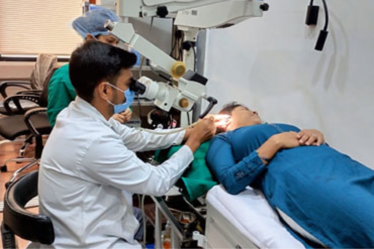
Ear microscopy is a specialized diagnostic procedure used to examine the structures of the ear—especially the ear canal and eardrum—under high magnification. It provides a clear, well-illuminated, and enlarged view of the ear, allowing for more accurate diagnosis and precise treatment compared to traditional handheld otoscopes. This procedure is commonly performed using a binocular operating microscope, which enables both hands-free examination and delicate microsurgical procedures when needed.
This technique is especially useful in evaluating conditions such as chronic ear infections, perforations of the eardrum, ear discharge, wax impaction, and the presence of foreign bodies. It is also an essential tool in pre-surgical assessments, postoperative follow-ups, and in performing procedures like suction cleaning, myringotomy, and removal of debris or infected material from the ear canal.
Ear microscopy is safe, quick, and typically painless. It is suitable for both adults and children and is usually done in a comfortable outpatient setting. The clarity and precision it provides ensure that even subtle changes or hidden issues can be detected early, allowing for timely and effective treatment. For anyone experiencing ear discomfort, hearing loss, or persistent infections, ear microscopy offers a deeper and more detailed insight into ear health, contributing to better outcomes and long-term care.
Schedule your consultation today and start your journey to better health!