


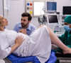


Otoendoscopy is a modern diagnostic procedure used by ENT specialists to examine the ear canal and eardrum with high precision. At Bhavik ENT Care, we offer advanced Otoendoscopy in Mumbai using specialized endoscopic equipment that allows detailed visualization of the ear structures. This procedure helps doctors accurately diagnose various ear conditions and provide effective treatment for patients experiencing ear-related problems.
Otoendoscopy is a safe, minimally invasive, and highly effective diagnostic method that plays an important role in modern ENT practice.
Otoendoscopy is a medical examination technique that uses a thin endoscope equipped with a camera and light source to view the ear canal and eardrum. The endoscope provides magnified images on a monitor, allowing the ENT doctor to carefully examine internal ear structures.
Compared to traditional otoscopy, otoendoscopy offers:
Better visualization
Higher magnification
Improved diagnostic accuracy
This helps ENT specialists detect even minor abnormalities within the ear.
Otoendoscopy is commonly performed to diagnose and evaluate several ear conditions such as:
Ear infections
Ear wax blockage
Eardrum perforation
Chronic ear discharge
Foreign bodies in the ear
Middle ear diseases
Cholesteatoma
Hearing-related ear problems
Early detection of these conditions allows doctors to provide timely and effective treatment.
Otoendoscopy provides several advantages compared to traditional ear examination techniques.
Key benefits include:
Clear and magnified visualization of the ear canal
Accurate diagnosis of ear disorders
Quick and painless procedure
Minimally invasive examination
Helps guide minor ENT procedures
Suitable for both adults and children
This technology significantly improves diagnostic precision in ENT care.
The otoendoscopy procedure is simple, safe, and usually takes only a few minutes.
During the examination:
The patient is seated comfortably.
A thin endoscope with a camera is gently inserted into the ear canal.
The internal ear structures are displayed on a monitor.
The ENT doctor carefully examines the ear canal and eardrum.
If required, images or videos may be recorded for diagnosis and treatment planning.
The procedure is generally painless and does not require anesthesia.
Otoendoscopy helps ENT specialists diagnose several ear conditions including:
Otitis media (middle ear infection)
Otitis externa (ear canal infection)
Eardrum perforation
Earwax accumulation
Cholesteatoma
Foreign bodies in the ear
Ear trauma or injury
Early diagnosis allows timely treatment and prevents complications.
Bhavik ENT Care is a trusted ENT clinic providing advanced ear diagnostic services.
Reasons patients choose us:
Experienced ENT specialists
Advanced endoscopic diagnostic equipment
Accurate and safe ear examination
Personalized patient care
Hygienic and modern clinical environment
Affordable consultation and treatment
Our goal is to provide precise diagnosis and effective treatment for all ear-related conditions.
If you are experiencing ear discomfort, hearing issues, or recurring ear infections, consult our ENT specialists at Bhavik ENT Care. Our clinic offers advanced Otoendoscopy in Mumbai for accurate diagnosis and effective treatment.
Book an appointment today and ensure the best care for your ear health.
Bhavik ENT Care is a specialized ENT clinic in Mumbai providing expert diagnosis and treatment for ear, nose, and throat conditions with advanced medical care and patient-focused services.
| Monday - Saturday: | 9:00 am - 10:00 pm |
| Sunday: | Closed |


Designee by Marketing Team – Shad & Karan